The Squat Test – Assessment and Correction of Movement Dysfunction
The squat is a basic, fundamental movement pattern that involves the lowering and raising of our body weight towards and away from the ground. This is a complex, multi-joint movement that involves adequate strength, flexibility, and coordination at the foot, ankle, knee, hip, and pelvis. The squat pattern is a valuable assessment tool for uncovering biomechanical problems that can be a causative factor in, or may predispose someone to lower extremity pain and injury. Correcting areas of dysfunction and rebuilding a proper squat pattern is an important component in injury rehabilitation, prevention, and ultimately, for improved movement efficiency and performance. Contact Kinetesis Spine & Joint Clinic today.
The remainder of this article will look more in depth at the squat test. Topics will include: 1) What constitutes a ‘normal’ squat test; 2) What are the most common dysfunctional patterns seen with a faulty squattest, including the causes of these patterns; and, 3) What are the best ways to correct a faulty squat test.
The ‘Normal’ Squat Pattern – What Should a Squat Look Like
To initiate the squat the ankle will dorsiflex, which allows the lower leg to rotate forward and the knee to translate forward over the toes. As this happens the knee and hip will flex and the trunk will adapt a slightly forward inclination, further accentuating flexion at the hip and helping to the keep the body’s centre of gravity balanced over the toes.
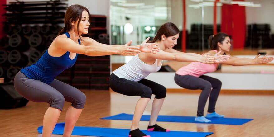
At the end position of the squat, when looking from the lateral perspective (sagittal plane assessment), with the feet flat on the ground the thighs should be in a horizontal position, the knees should be positioned directly over over the toes, and the trunk and lower legs should both be inclined forward and should be parallel to each other. With respect to the trunk, the lower back should maintain a slight inward lordotic curve and the thoracic spine should bend slightly into extension. This thoracic extension is usually seen as a slight flattening out of the middle/upper back). (Note: for a deeper squat the curve in the lower back will reverse directions, but this should not happen until deeper into the squat. This puts the lower back in a vulnerable position so care must be given if using a deep squat as a training exercise).
When looking from the front (assessing the frontal and transverse planes) the critical assessment point is to determine if the ankle, knee, and hip remain stacked over each other. Remember, the squat is primarily a sagittal plane motion (i.e., involves motion only in the front-to-back direction). Any side-to-side or twisting movements during the squat is considered dysfunctional. (Note: while technically speaking there are some very subtle rotations that occur at the knee and hip, and there is some tri-planar motion at the ankle, the magnitude of these motions is very small and is visually discernible to only the most skilled observer. So, for all intents and purposes, consider the squat a unidirectional motion).
Determining if the lower extremity is held in the correct alignment is most easily assessed by looking at the angle formed by 1) the thigh and lower leg, and 2) the lower leg and the foot. Essentially, the alignment of the hip, knee, ankle, and foot observed during a relaxed, standing position should be maintained during the squat. Additionally, the pelvis should be observed to determine if there is any twisting or side-to-side deviation as the body is lowered towards the ground. This would indicate an asymmetry within the squat pattern that likely requires further investigation.
The Role of the Muscular System
While adequate flexibility at the hip, knee, and ankle is a key requirement for a proper squat (restricted motion at any of these joints will block the normal squat motion) adequate muscle strength and coordination is equally important. Muscle action during the squat can be divided into two roles: 1) muscle force required to generate movement; and 2) muscle force required to stabilize the lower extremity and and maintain the alignment between the hip, knee, ankle, and foot.
The key movement muscles are the superficial calf muscles (gastrocnemius and solues), the quadriceps, and gluteus maximus. These muscles must act in the sagittal plane (i.e., the front to back direction) to flex and extend the ankle, knee, and hip as the body is raised and lowered to the ground. Problems with these muscles will lead to dysfunctional movements in the sagittal plane, and are evident when observing the squat from the side (see below).
The key stabilization muscles are the muscles of the foot and deep calf muscles (tibialis posterior, flexor hallicus longus, flexor digitorum longus), the lateral hip muscles (gluteus medius and minimus), and deep hip external rotator group. These muscles act in the frontal and/or transverse plane, and must work to prevent excessive motion of the foot, lower leg, or thigh in the rotational or side-to-side direction. Problems with these muscles are evident when observing the squat from the front (see below).
Recognizing Dysfunction in the Squat Pattern
The Lateral View
When assessing the Squat from a lateral view, one of the most common problems seen is a position in which the knees pass too far in front of the toes at the bottom of the motion. This is often referred to as a patello-femoral shear pattern. This is often seen in conjunction with the trunk staying too upright so the that lower legs are inclined much farther forward compared to the trunk (remember, the trunk and lower legs should be parallel). With this position the body is lowered more from the knee and ankle (these joints move too much), while the hip motion and force contribution is that under normal circumstances (this hip does not flex enough). Interestingly, many people think squatting this way is better, possibly due to the common advice of “lift with your legs, not with your back”. While this pattern does in fact take stress off the back, the load is instead re-distributed to the ankle, and even more so to the knee, causing the muscles surrounding these joints (the quadriceps and calf/achilles) to supply more of the muscle force needed to raise and lower the body in our out of the squat. In fact, this this pattern is very common in people who suffer from anterior knee pain/ patellofemoral syndrome, as well as achilles tendinopathy. Common causes of this pattern often include a lack of flexibility of hip flexion or internal rotation, weak hip extensors, and sometimes weak trunk extensors (the body avoids forward inclination of the trunk to protect the back).
While less common, the opposite pattern in which the trunk flexes too far forward while the knee and ankle demonstrate reduced motion can also be seen. In this pattern the trunk incline angle is greater than that of the lower leg. Similar to the pattern described above, the areas of the body that move more than normal, in this case the hip and/or back, are doing more of the work. This makes them more susceptible to pain and overload, while the knee and ankle contribute less muscle force compared to normal. Common causes of this pattern include ankle dorsiflexion restriction, ankle/calf weakness, or quadriceps weakness.
The Anterior View
The most common dysfunction pattern seen from the anterior view is the thigh turning inward as the body is lowered towards the ground (can be one or both knees). This pattern is usually caused by a weakness in the hip abductor and external rotator muscles (these muscle are located on the back of the hip and include the Gluteus Maximus and Minimus, Piriformis, Inferior and Superior Gemelli, and Obturator Internus). These muscles must contract to stabilize the hip joint and prevent the thigh from rotating inward as the body is lowered down. When there is a problem with any of these muscles the thigh will fall inwards. Because the foot is fixed on the ground the lower leg will be pulled along with the thigh. This in turn puts a lot of strain on the knee and can lead to pain on the sides of the knee. (The knee is great at bending forwards and backwards, but does not like to twist. Twisting of the knee will stress the meniscus, medial or lateral joint capsule, medial collateral ligament, iliotibial band,and pes anserine bursa).
This pattern can also be caused by a problem at the foot and ankle. When the foot is involved the foot can also be seen to fall inwards and the arch will often flatten out (this is referred to as hyperpronation of the foot). As the foot falls in it will pull the lower leg with it, and the lower leg will in turn pull the thigh inwards as well. Foot hyperpronation can be related to structural issues in the feet (i.e. how the bones were formed during development), but can also be related to restrictions in the muscles and ligaments around the ankle or weakness in the deep compartment of the calf (these muscles include the tibialis posterior, flexor digitorum longus, and flexor hallicus longus).
Because of the interconnectedness of the lower extremity It is not uncommon for both of these problems to co-exist. In my experience, it is more often the hip that is the primary dysfunction and correction of the hip often will alleviate both problems.
Correcting Dysfunctional Movement Patterns
It is important we are able to perform functional movements such as the squat pattern properly. Remember, as the squat pattern is repeated many times per day a dysfunctional pattern can easily lead to the accumulation of micro-trauma and eventually pain and injury.
The key to correcting the squat is identifying the specific muscle and/or joint problems at the foot, ankle, knee, hip (especially the hip), and first addressing those problems. In some cases this can often be done with specific stretches or exercises. In other cases the muscles and joints may be contain scar tissue / soft tissue adhesions. When this is the case these adhesions need to be released before the tissues will respond to stretches and exercises. Active Release Techniques (ART) treatment works well for this (see our article on ART for more information). As the local problems are resolved and the hip, knee, and foot can all work the way they are supposed to, you can then start to train the squat into your rehabilitation or fitness routine, being careful to use proper form. Feel free to contact our physiotherapist located at Bedford & Fall River, NS.